Orthopedic procedures have evolved dramatically over the past few decades. One major advancement is arthroscopy. This technique allows doctors to enter joints with minimal disruption. Surgeons make a tiny incision and insert a camera and tools. The camera projects images onto a screen during the procedure. This provides a full view of the joint’s internal structure. The procedure doesn’t require large incisions or extended recovery time. Arthroscopy became a standard option for joint diagnostics and minor repairs.
Patients often experience less postoperative pain with arthroscopy
Open joint surgeries typically require long healing periods. The surrounding tissues are heavily affected during traditional procedures. Arthroscopy avoids most of this trauma. Because incisions are smaller, there’s less blood loss. Patients usually go home the same day. Pain is milder, and many return to activity sooner. This benefit makes arthroscopy especially attractive for athletes and active individuals.
Damage to cartilage can be repaired without cutting open the entire joint
Joint pain often stems from cartilage tears. These tears may not heal on their own. In the past, open surgery was the only repair option. But now, surgeons access the damaged area with precision tools. The joint remains largely intact. Small shavers and sutures are used to reshape or reattach tissue. Recovery time is shorter than traditional methods. Patients also report better joint function after surgery.
Diagnostic arthroscopy helps confirm what imaging can’t reveal
MRIs and CT scans provide important details. But they don’t always capture subtle tissue injuries. Arthroscopy offers direct visual access. Surgeons can inspect ligaments, cartilage, and the joint capsule. This real-time assessment clarifies uncertain diagnoses. Some conditions only become visible inside the joint. It helps doctors make immediate decisions during the procedure. That means fewer follow-up surgeries.
Shoulder, knee, hip, and wrist joints are the most common arthroscopy sites
Each joint has unique challenges. But some areas are particularly suited to arthroscopic treatment. The knee is the most common site, especially for meniscus tears. The shoulder follows closely, due to rotator cuff injuries. Hip and wrist procedures are more specialized. These areas require customized instruments due to tighter spaces. Surgeons train extensively in joint-specific techniques to perform them correctly.
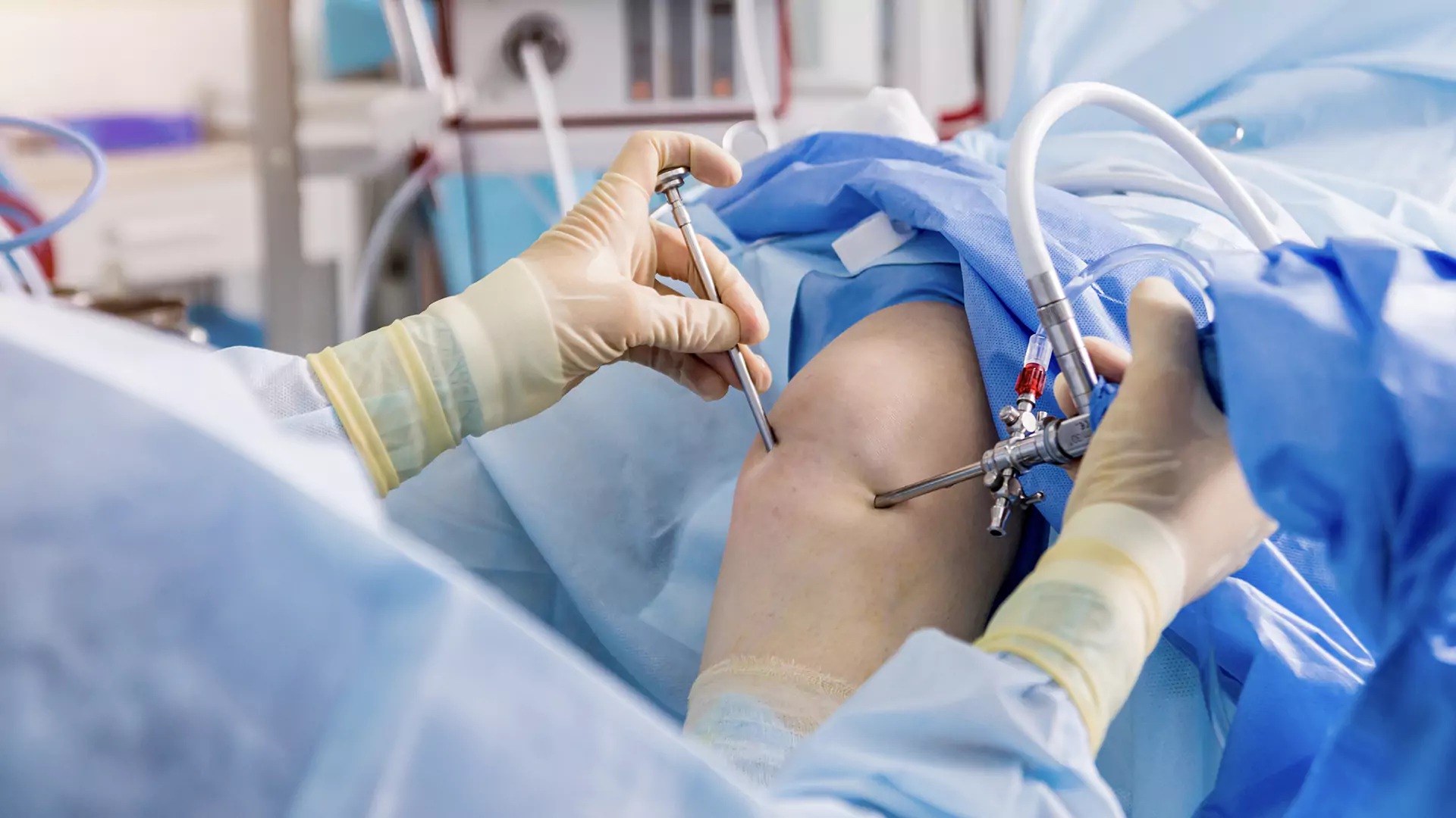
The arthroscope projects images from inside the joint onto a high-definition screen
The camera at the tip of the arthroscope is small but powerful. It sends images to a connected monitor. Surgeons rely entirely on this video feed during surgery. It offers detailed views of even the tiniest structures. High-definition quality is essential for precise movements. The setup turns a hidden joint cavity into a clearly visible space. This transformation allows accurate, controlled actions.
Movement is often restored more quickly than with traditional joint operations
Rehabilitation begins sooner after arthroscopic procedures. Patients can start light movements within days. Physical therapy focuses on restoring flexibility and strength. Swelling is lower than in open surgeries. This promotes faster joint function recovery. Most patients return to regular activity in a few weeks. Full recovery still depends on injury type and patient discipline.
Arthroscopy carries risks like bleeding, infection, and nerve injury
Despite its minimally invasive nature, arthroscopy isn’t risk-free. Every surgical procedure involves potential complications. Some patients experience bleeding within the joint. Infections may occur if proper hygiene isn’t maintained. Nerve damage is rare but can cause lingering numbness or pain. Surgeons explain these risks before any intervention. Proper technique and sterile environments reduce their likelihood significantly.
The decision to undergo arthroscopy depends on injury severity and lifestyle
Not all joint problems need surgery. Some can be managed with therapy or medication. Arthroscopy is recommended when symptoms don’t improve. It’s also preferred when joint function limits daily life. Athletes often choose it to speed up return to play. Others prefer conservative care unless absolutely necessary. Shared decision-making with a specialist is key.
Recovery timelines vary based on procedure complexity
A simple joint inspection might require only a week of rest. Repairing torn cartilage may need several weeks. Ligament reconstructions take longer, often months. Surgeons provide individual recovery plans after the procedure. Follow-up visits monitor progress and prevent complications. Adherence to instructions plays a major role in success.
Local or general anesthesia is used depending on the procedure and joint involved
Smaller joints like the wrist or ankle may require only local anesthesia. Larger procedures often use general anesthesia. The choice depends on patient health, pain tolerance, and procedure duration. Anesthesiologists assess risk factors before surgery. Many arthroscopies now take less than an hour. Outpatient status means patients return home the same day.
Patients are advised to avoid high-impact activities during early recovery
Returning to sports too soon risks reinjury. Joint tissues remain vulnerable during early healing. High-impact movements stress the healing area. Surgeons usually restrict running or jumping for weeks. Patients can walk, stretch, or swim with guidance. Strength training resumes gradually. Each step is monitored to prevent setbacks.
Small scars and minimal muscle disruption make arthroscopy appealing
One visible advantage is the cosmetic outcome. Arthroscopy leaves only small scars. These usually fade within months. Muscle tissues are not cut, just bypassed. This allows better strength retention post-op. Cosmetic results matter for some patients, especially younger individuals. Others appreciate less visible evidence of surgery.
Technological advances have made tools more precise and procedures shorter
Early arthroscopes were bulkier and had limited vision. Modern tools are slimmer and sharper. Camera clarity has improved dramatically. Robotic arms and 3D navigation support complex cases. These developments reduce surgery time and improve accuracy. Patients benefit from fewer complications and faster recovery. Surgeons gain confidence from better visibility and control.
Insurance coverage for arthroscopy depends on the indication and policy details
Medical necessity determines insurance approval. Some providers require imaging before approving surgery. Sports injuries often meet the criteria quickly. Chronic joint pain without imaging support may be denied. Patients should check pre-authorization requirements in advance. Policies vary between providers and countries. Documentation and doctor’s notes help support approval.
Training in arthroscopy requires precision and visual-spatial coordination
Not all orthopedic surgeons are arthroscopy specialists. It requires specific training beyond medical school. Surgeons must develop excellent hand-eye coordination. They practice in simulators before operating on patients. Workshops and fellowships focus on joint-specific techniques. Skills are refined continuously to adapt to new tools. Confidence grows with each successful case.
Elderly patients benefit from shorter anesthesia and less surgical trauma
Older adults often face higher surgical risks. Arthroscopy offers a safer alternative in many cases. Shorter procedures mean less exposure to anesthesia. Smaller incisions lower infection and bleeding risks. Post-op pain is easier to manage. These advantages reduce hospital stays. Geriatric patients often prefer this route for joint issues.
Joint injuries in younger patients are often treated arthroscopically
Children and teens frequently experience sports-related joint problems. Torn meniscus or dislocated shoulders are common. Arthroscopy avoids growth plate damage. Recovery is usually quicker than with open surgery. Parents often choose this option to minimize long-term effects. Pediatric specialists tailor each approach to age and anatomy.
Sterile fluid is used to expand the joint space during the procedure
During arthroscopy, sterile fluid is pumped into the joint. This creates room for instruments to move. The expanded space improves visibility and reduces the chance of tissue damage. After the procedure, the fluid is drained. Some swelling remains temporarily. It subsides within a few days. Proper fluid management is part of surgical safety.